Share OCT and fundus imaging
Studies, images and reports can be released digitally for follow-up care or clinic access.
PACSY makes ophthalmic imaging securely available to patients, clinics and care partners as a simple extension to existing systems.
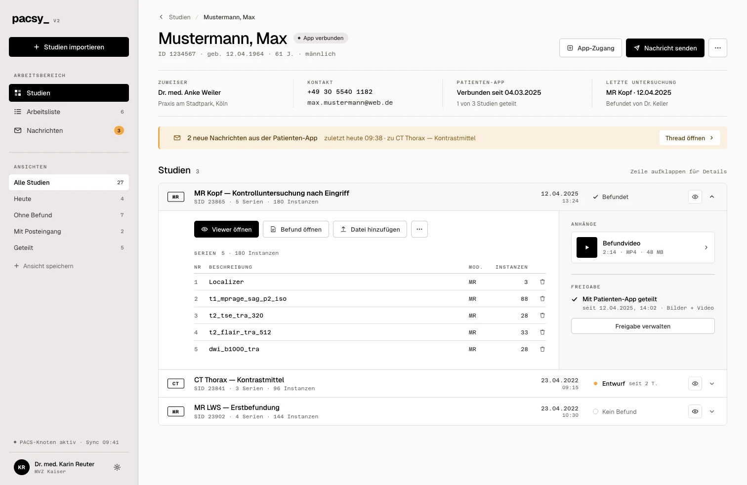 Product preview with sample data
For OCT, fundus imaging and follow-up, comparability matters. PACSY helps share relevant data securely without patients physically transporting records.
Studies, images and reports can be released digitally for follow-up care or clinic access.
Control examinations stay easier for providers to access and compare.
Patients can take relevant imaging data with them without requesting printouts or discs.
Less manual exporting, less media handling and fewer questions about missing images.
Make control examinations digitally available for retinal and macular follow-up.
Provide imaging securely for follow-up care or second opinions.
Share relevant data for procedures, referrals and postoperative controls.
Medical images need clear responsibilities. For storage, access and the use of the medical viewer.
Explore PACSY around your practice, your systems and your team.
Your contact